Pages 62-73 (including rear covers)
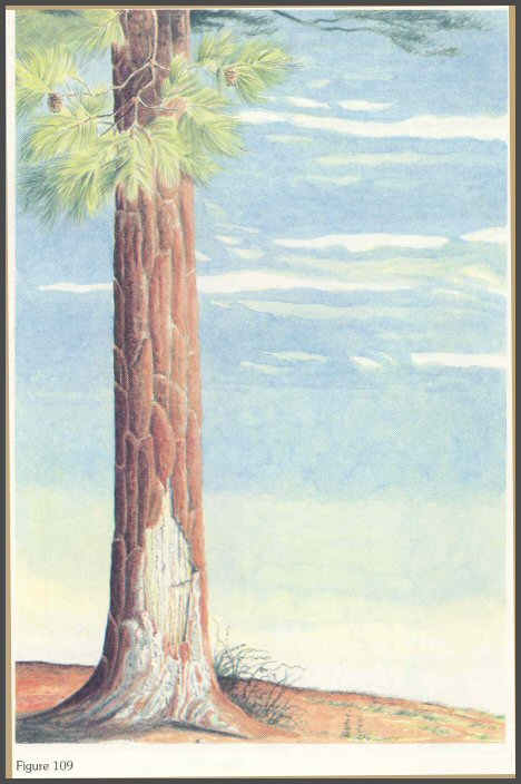
A large basal wound after a few weeks on a pine. (fig. 109)
Page 62
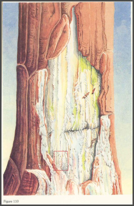
A copious flow of resinous material is obvious on the wound surface. (fig. 110)
Page 63
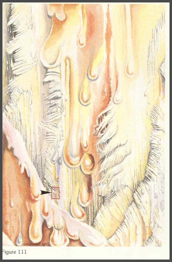
The torn wood, debris, and droplets of moisture and resin become visible at this magnification. (fig. 111)
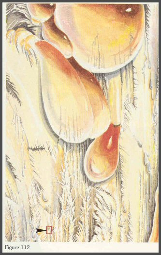
Here the droplets act as prisms that reflect the light like minute rainbows. The minute wood splinters now appear as gigantic spears coated with multicolored resin. (fig. 112)
Page 64
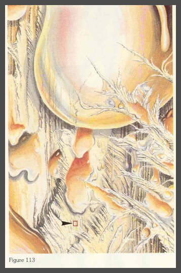
Droplets and torn wood not visible to the human eye now become visible. This is the view small insects have of the wound. (fig. 113)
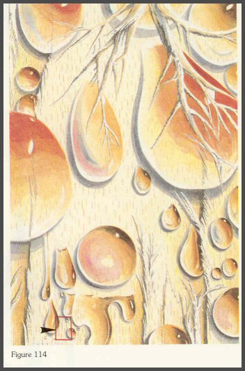
Here the tangential ends of the rays begin to show. The minute splinters appear as gigantic branches that trap the minute droplets of moisture and resin. Each droplet supports many colonies of competing microorganisms. (fig. 114)
Page 65
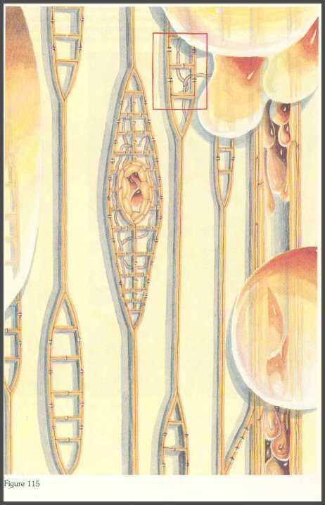
Several weeks later, the fungi and bacteria have begun to infect the wood. A diagrammatic view shows the ray cells infected, and a resin pocket forming as the walls of the surrounding cells begin to dissolve. (fig. 115)
Page 66
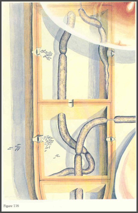
A blue-stain fungus and many bacteria in ray cells. The bacteria are clustered near the pits. The microorganisms begin to digest the cell contents, and blue stain results. (fig. 116)
Page 67
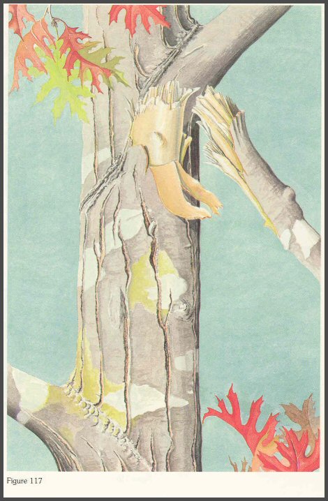
Broken and dying branches are some of the most common infection courts for microorganisms. Large branch stubs on trees (that have heartwood) expose heartwood. (fig. 117)
Page 68
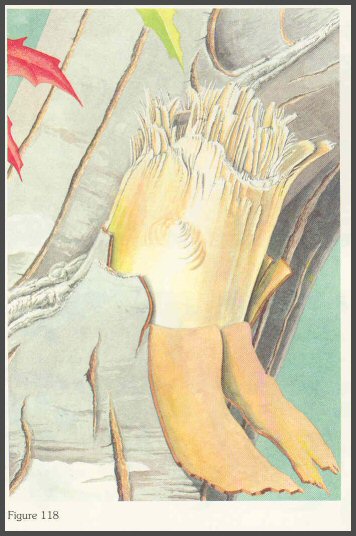
A closer view of a branch stub on an old oak. (fig. 118)
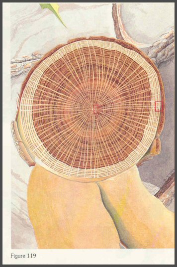
(Below) When the stub surface is sawn smooth, this is the view of the old branch with central heartwood. (fig. 119)
Page 69
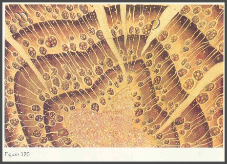
A closer view of the heartwood near the branch center shows the vessels plugged with tyloses. (fig. 120)
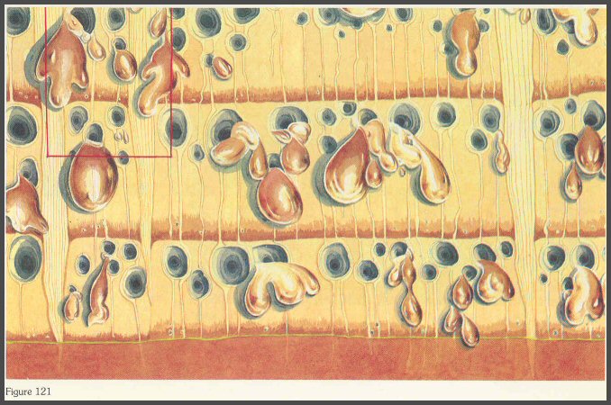
(Below) A closer view of the sapwood shows the open vessels oozing moisture. (fig. 121)
Page 70
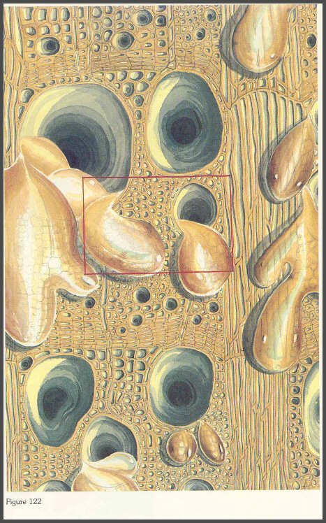
The vessels appear as large tunnels or caves at this magnification. (fig. 122)
Page 71
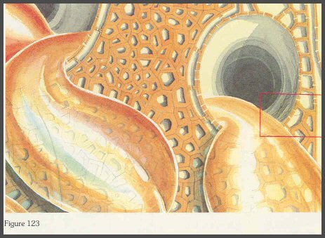
The ordered pattern of fibers surrounding the vessels is shown here. Each microscopic droplet of moisture could be the site or battleground for intense competition by many groups of different microorganisms. (fig. 123)
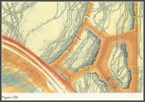
After several weeks, the fungi and bacteria invade. The small dots are the bacteria. The cells have been injured or killed, so invasion is an easy matter for the microorganisms. They reproduce rapidly and begin to interact with the tree. (fig. 124)
Page 72
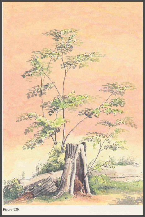
In the end, trees will continue to grow, die, and decay; and grow, die, and decay; and grow, die, and decay. (fig. 125)
COMPARTMENTALIZATION extends the time a tree will grow under the constant stress of wounds.
SUCCESSIONS make it possible for microorganisms to survive against the protective forces of a tree.
AN UNDERSTANDING OF THE EXPANDED CONCEPT OF DECAY GIVES US BETTER OPPORTUNITIES TO REGULATE TREE DECAY.
The End