[Pages 1 to 31]
The guide gives an historical pictorial overview of the development of the CODIT model. CODIT is a model of Compartmentalization Of Decay In Trees. CODIT shows how trees are built up in highly ordered compartments. Trees survive after injury and infection so long as they recognize and compartmentalize the injured tissues, confining them to small volumes rapidly and effectively. When trees break down from disease or decay, they do so compartment by compartment.
Field dissections that led to the CODIT model are shown first. The application of the model to many other tree problems is then shown. A constant thread that runs through the photos is that many problems in wood products have their origin in the living tree.
Page 1
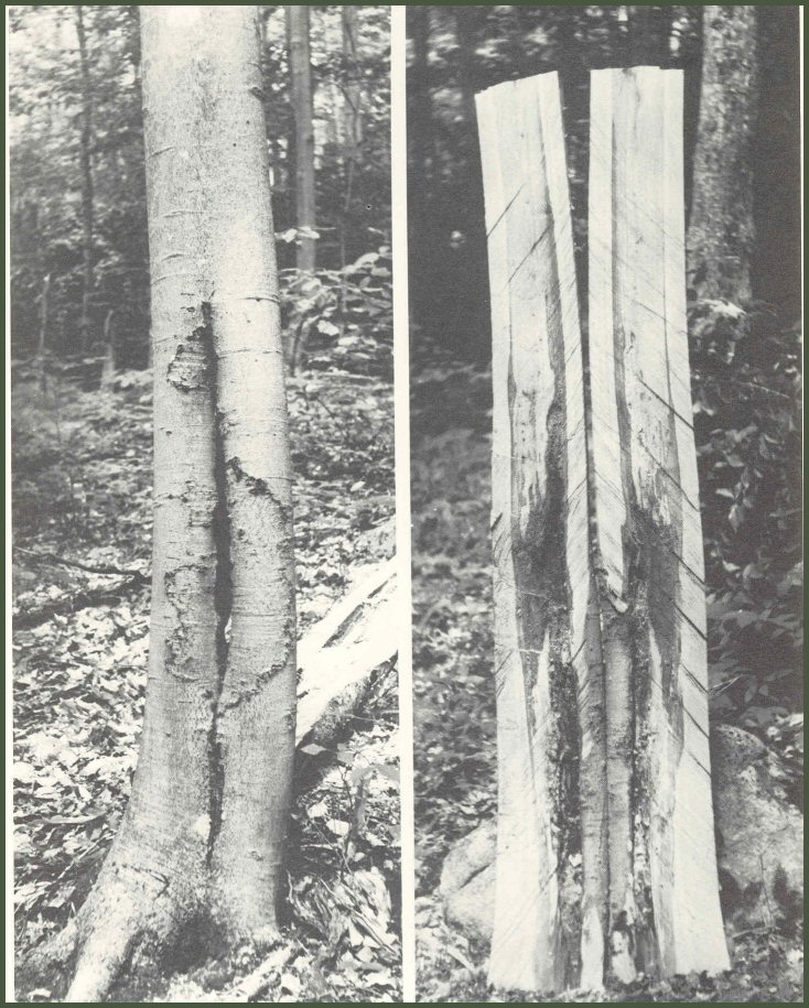
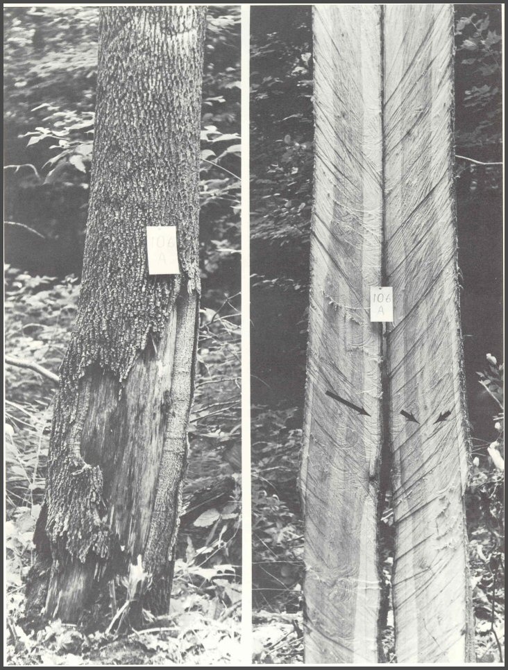
1. Thousands of trees with wounds were dissected longitudinally. The decay did not spread outward into new wood formed after wounding, as this American beech shows.
Page 2-3
2. And this beech with decay associated with Fomes igniarius.
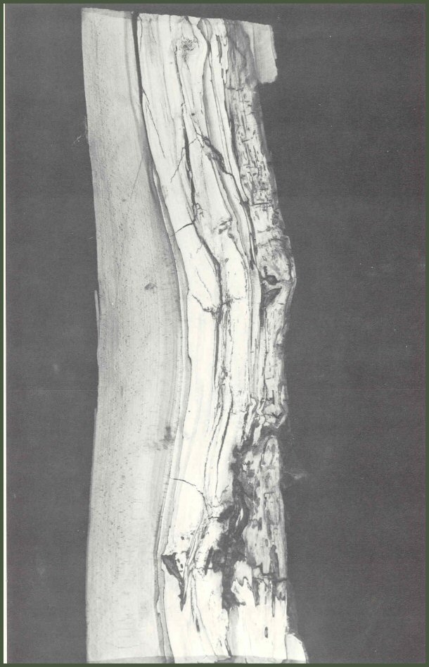
Page 4-5
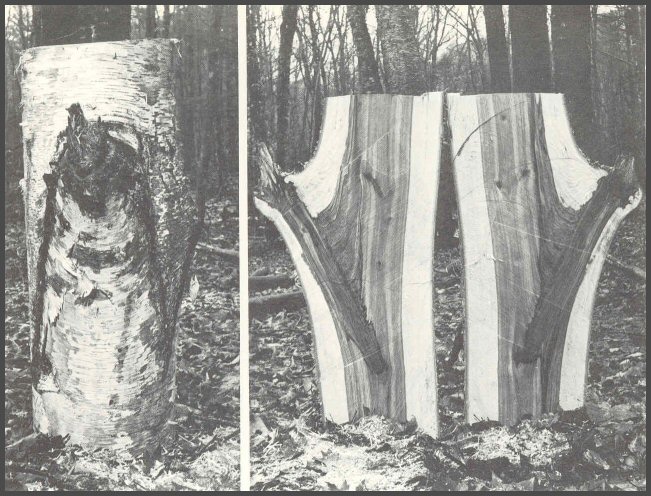
3. Cross-sections of trees showed the same patterns: the decay was confined to the wood present at the time of wounding, as in this yellow birch.
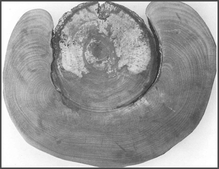
Page 6-7
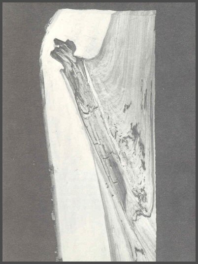
4. New questions arose as more trees were dissected. Dissection of this paper birch showed that the central column of colored wood was associated with the wounds.
Most birch trees did have a central core of colored wood, but some trees, even large, old trees, had no colored cores. The discolored wood was associated with wounds.
Dissections showed that some columns of discolored wood associated with the wounds were very small, while others were large. Wound size and depth were important factors affecting size of internal columns of discolored and decayed wood.
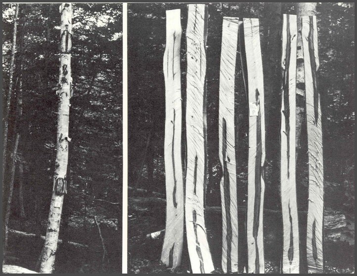
Page 8-9
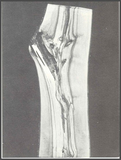
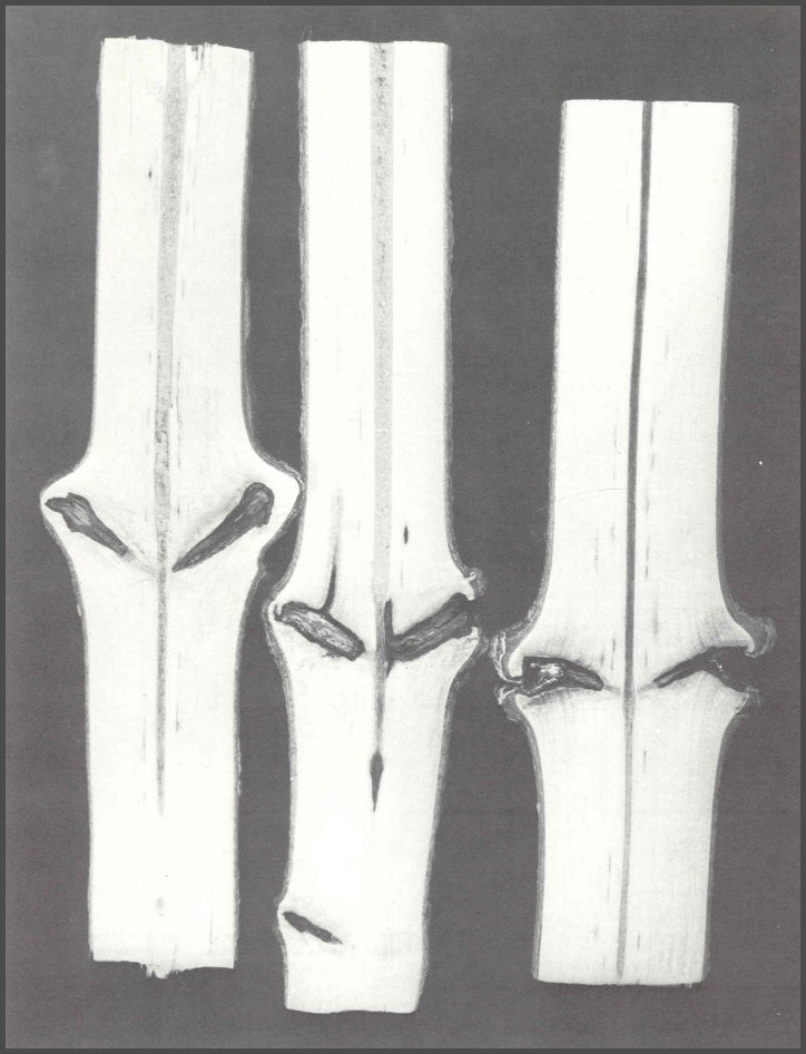
5. The wound on this yellow birch was 50 years old. The large arrows show the size of the discolored core at the time of the large wound. The small arrows show the size of the tree at the time of wounding. The decay did not spread inward into the wood that had been altered by previous wounds, or outward into the new wood that formed after wounding.
Page 10-11
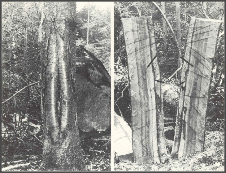
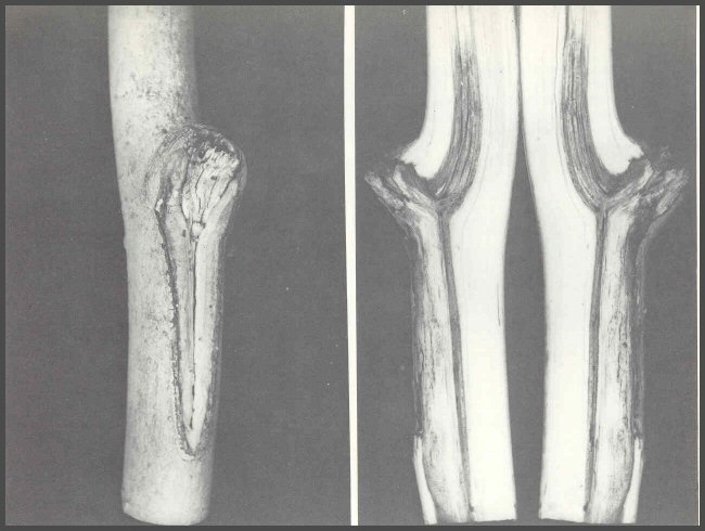
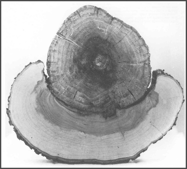
6. Patterns were complex in sugar maple, but more dissections revealed their high order. This tree had an old basal wound-over 75 years old. The fruit body was Fomes connatus. The large wound above was inflicted by sugar maple borers, Glycobius speciosus. The type of decay associated with each wound was different, but the fungi were confined to the wood present at the time of wounding, even after more than 75 years.
Page 12-13
7. More and more sugar maple trees with clear wood to the pith were found. The 8-year-old basal wound caused very little defect. A dark line was often seen in the growth ring that formed after wounding (arrow). The question of whether beech, birch, and maple have a normal central core of colored heartwood began to emerge.
8. The dark line (arrow), was first called "barrier cells". The cells formed after wounding, and they separated the infected wood from the sound wood. Barrier zone was the term given later to these cells. The barrier zone was easy to see in sugar maple. The pattern of discolored wood forming first, and then decayed wood spreading within the discolored wood, and the pattern of bacteria, non-decay-causing fungi, and decay- causing fungi all began to take shape.
Page 14-15
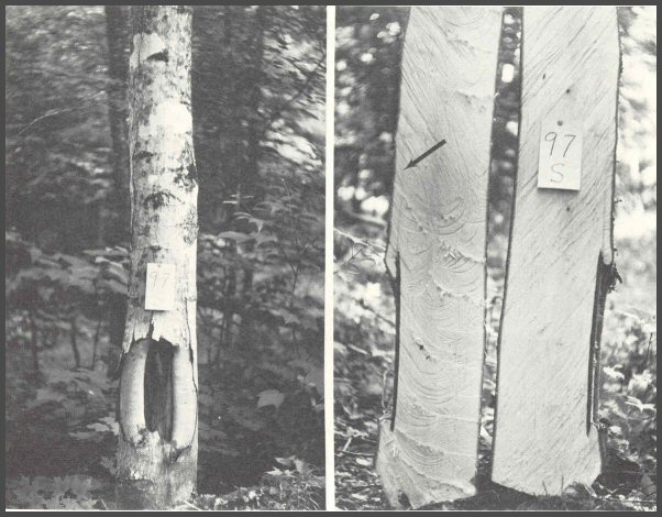
9. Dissections of American ash showed the same internal patterns as the other species of hardwoods. Note the light colored central column (small arrows). This was associated with many small stubs. The large arrow shows a barrier zone.
Page 16-17
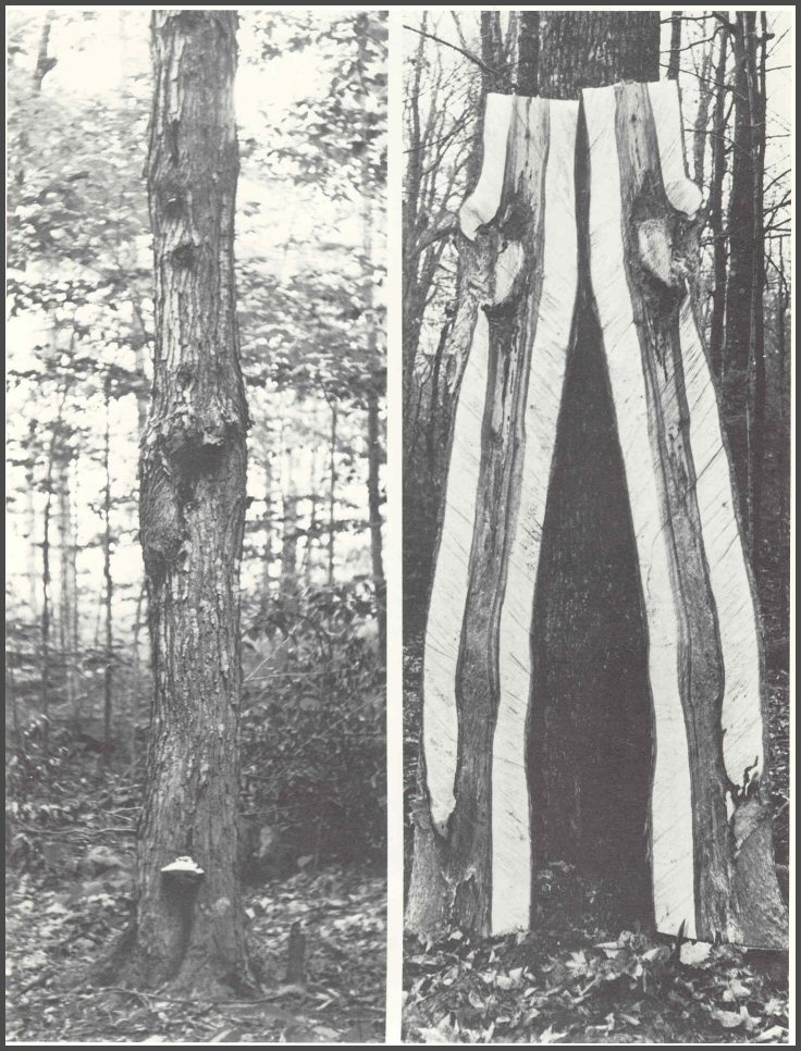
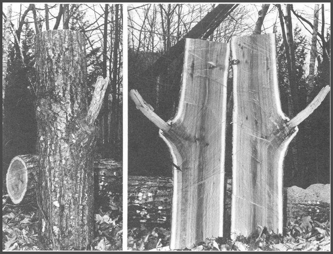
10. Dissections began to focus on branch stubs. Decayed wood associated with large old stubs was confined to the wood present at the time the branch died. Note that the discolored wood associated with the stub on this paper birch does not spread into the central column of discolored wood associated with older branch stubs.
11. This large old stub on a paper birch was "pinched off". The decayed wood is strongly walled off within the old internal branch wood. The decay did not spread outward into the healthy wood.
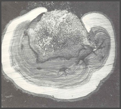
12. The hollow branch base in this yellow birch was surrounded by a light- colored column of decayed wood. The central column of decayed wood was associated with many branch stubs. Each new column of decayed wood remains separate from the others. This pattern would be impossible to understand from cross sections.
Page 18-19
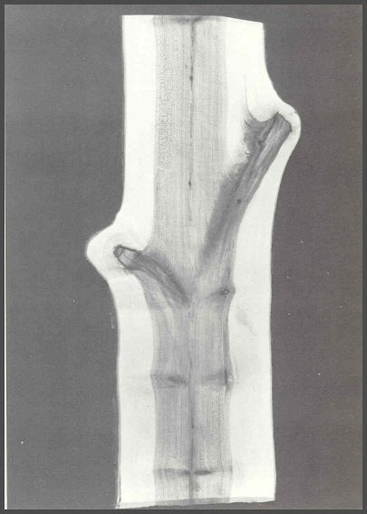
13. The dead spot below this small stub on a red maple indicates that the cambium was injured when the branch tore away from the main stem. The discolored wood was walled off within the wood present at the time the branch died.
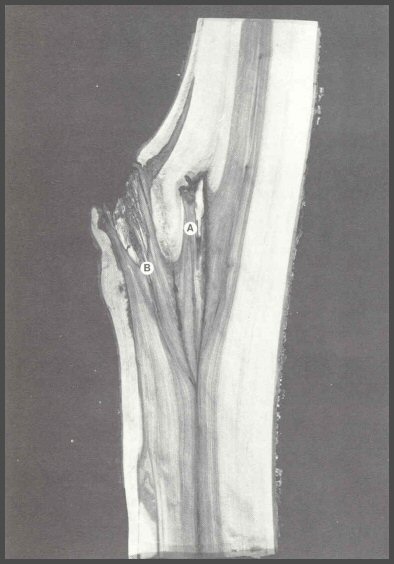
14. Two major leaders died on this yellow birch, and the third leader grew to be the main stem. Note how neatly the leaders are walled off from each other. The decayed socket in stub A was essential for effective shedding. A similar socket of decayed wood is in stub B. The upward extension of discolored wood associated with stub A indicates that the trunk was injured when stub A fell away from the trunk.
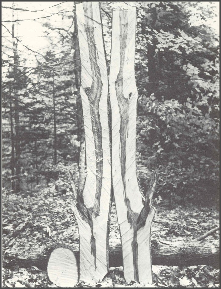
15. Two large old branch cores on red maple. Note the tip of decayed wood in the lower branch core. Decayed wood is essential to shedding. The decayed wood extended farther into the inner branch wood in the upper branch. The central core of discolored wood in beech, birch, maple, ash, and many other trees is associated with the death of branches and the alteration (and usually infection) of the wood present at the time the branch died.
Page 20-21
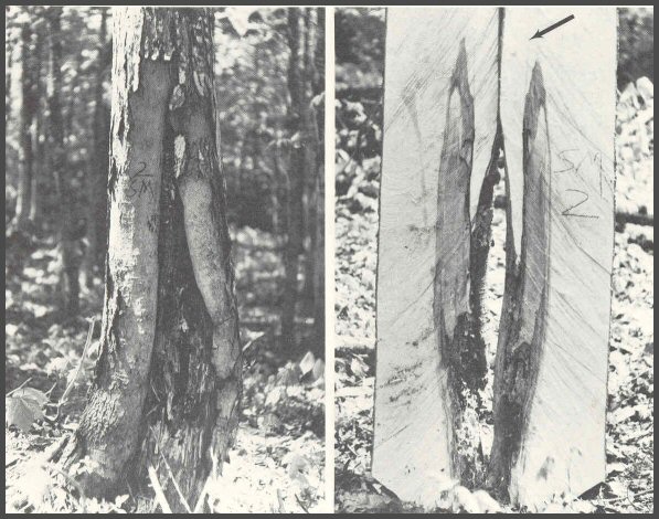
16. Dissection of this sugar maple shows a clear base, but discolored wood associated with branch stubs. The discolored wood in the trees studied yielded many species of bacteria and fungi, usually species in the genus Phialophora and closely related genera. The results began to show a succession of microorganisms associated with discolored and decayed wood.
Page 22-23
17. Dissection of very small stems showed patterns similar to those of large branches and similar microorganisms were isolated from the discolored wood. The small inner branch cores were walled off. It appeared also that there was a blocking of the pith connection between branches and main stem.
Page 24-25
18. Dissections of branch stubs on oaks and other heartwood-forming trees showed patterns similar to those in birch and maple. The discolored wood associated with the dead branch on this red oak was walled off within the wood present at the time the branch died. The discolored wood was discolored heartwood (arrows). Heartwood will wall off defects, and heartwood will discolor. If heartwood is an unresponsive tissue, how can this be?
19. It was time to begin rethinking our beliefs about heartwood. The classical concept of tree decay, the heartrot concept, would have you believe that once heartwood was infected, the fungi would grow at will into a hollow formed by complete digestion of heartwood. It was time to question this concept.
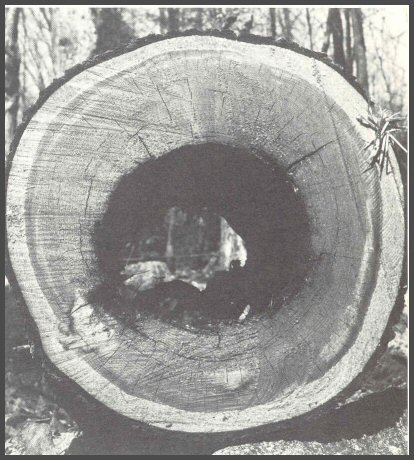
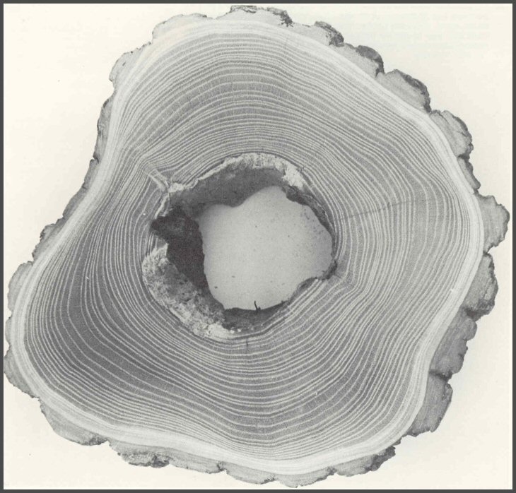
20. Dissection of white pines showed that the diameter of hollows was the diameter of the tree when it was wounded. Sound heartwood surrounded the hollow. How could this be so if the classical heartrot concept were true?
Page 26-27
21. Dissections of tree species that have a normal heartwood showed patterns similar to those in birch and maple. The decayed wood in this black cherry did not develop into the surrounding heartwood, even after 20 years. Where small radial cracks formed, the decayed wood did move out, but still only very slightly. It appeared that the heartwood- forming trees also had similar patterns of discolored and decayed wood, even within heartwood.
Page 28-29
22. Similar patterns were seen in other heartwood-forming trees such as this black locust. The diameter of the hollow was the diameter of the tree at the time of injury. Even after almost 40 years, the so-called "heartwood rotting fungi" did not spread outward into the heartwood that surrounded the hollow. These observations suggested that microorganisms could only infect wood that was first altered by injury.
Page 30-31