Page 51-75
[Note from the web master. The word heal may be used
here to describe the closure of a wound. Dr. Shigo now calls it closure and not
healing. That and other terms can be found at
www.treedictionary.com for your use.
John A. Keslick , Jr.]
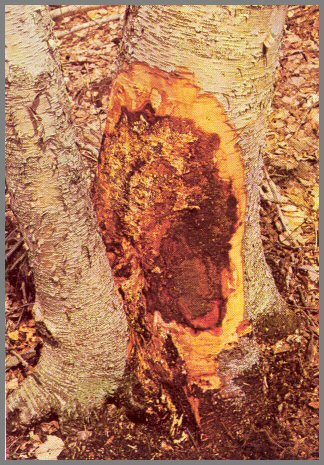
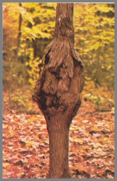
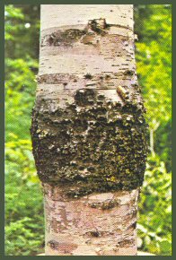
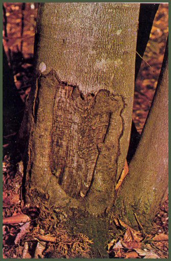
FIGURE 33. - FOMES CONNATUS FRUIT BODY AND BORER WOUND.
The large fruit body on the face of the old basal wound on this sugar maple is Fomes connatus. It is typically white and moist, and has green moss on top. Above it is a large open wound caused by a sugar maple borer. Borer wounds can be found on all parts of the stem. These beetles apparently are attracted to the trees left after logging. We do not know whether it is the open condition of the stand or the stress after logging that favors infestation.
Dissection shows that the sugar maple borer attacked the tree soon after it was wounded at the base. The decay due to F. connatus is dark brown to black, and moist. The decay is advanced, but ends a short distance above the fruit body. Another decay fungus is advancing downward from the borer wound. The diameter of the defect column indicates the diameter of the tree when it was wounded.
Page 51
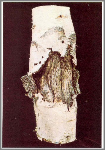
FIGURE 34. - ARMILLARIA MELLEA INFECTING BASE OF PAPER BIRCH
The wet orange-brown ooze at the base of this paper birch is a
good indicator of Armillaria mellea infection. Here the bark was
chipped away to show the dead area at the base of the birch and the plates of
white fungus tissue. This fungus usually advances upward from the roots.
Infected trees should be harvested as soon as possible.
This fungus may act either as a cambium killer, as shown here, or as a root and
butt rotter, as shown in the three photographs that follow.
Page 52
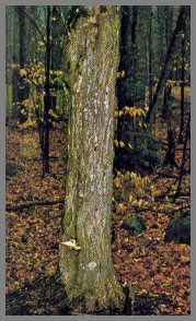
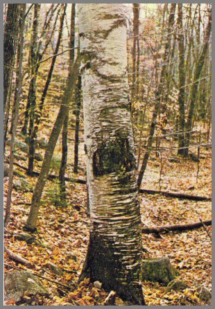
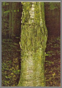
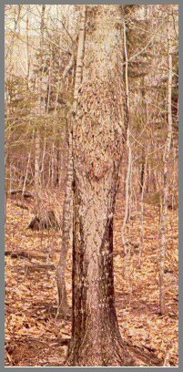
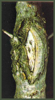
FIGURE 35. - COLLAR CRACK OF PAPER BIRCH.
In yellow birch and paper birch, Armillaria mellea infects the roots, and cracks form at the base of the trees. Once the cracks form, other decay fungi have easy access to the bases of the trees. Usually fungi that are more aggressive than A. mellea above the base enter at these openings. Once this occurs, the base decays fast. Such trees should be cut as soon as possible.
Page 53
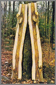
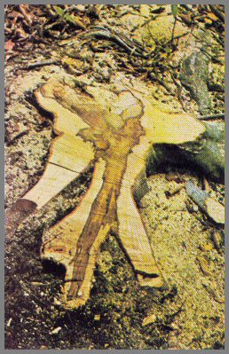
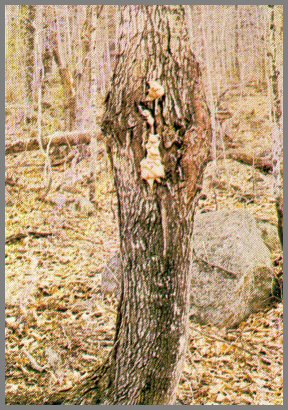
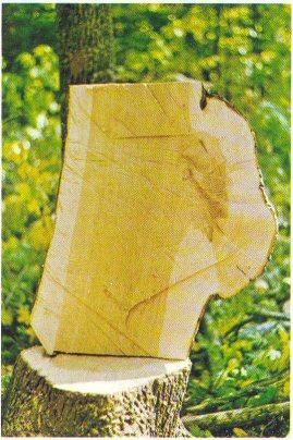
FIGURE 36.-HOLLOW BUTT CAUSED BY ARMILLARIA MELLEA.
Dissection of this beech tree shows a hollow butt (left) caused by Armillaria mellea advancing upward from the roots. The hollow in the upper section (right) was caused by other fungi advancing downward from branch stubs. If you see only the ends of logs, and both ends have hollow centers, you might guess that what you see is all one defect from one cause. Often this is not so. Butt decays often end abruptly in northern hardwoods.
Page 54
FIGURE 37.-ARMILLARIA MELLEA ROOT DECAY.
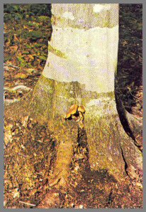
The fruit body of Armillaria mellea at the base of this beech tree
indicates root and butt decay. Fruit bodies like this can be found only in
the fall. Cracks on roots and wet spots usually indicate root decay.
Dissection shows that both the butt and root are decayed. In early stages
the infected wood is dark brown and very moist. Dark zone lines are common
in the dark discoloration and decay. In later stages A. mellea
decay is moist, bleached, and stringy. The fungus produces black ropy
strands of mycelium that resemble shoestrings - hence the name shoestring
fungus. Other organisms, especially bacteria, are associated intimately
with A. mellea.
Page 55
FIGURE 38.-NECTRIA CANKER ON PAPER BIRCH.
This target canker on a paper birch tree is caused by Nectria
galligena. Such cankers usually begin at branch stubs. The face
of the canker is hard and light in color. Very little discoloration is
associated with these cankers; it may go only a few inches above and below the
canker. The cankers themselves are highly decay resistant-except in trees
also infected with Poria obliqua. Sapsuckers often tap the stem
above the canker. These cankers are common on trees on poor sites,
especially dry mountain tops.
Page 56
FIGURE 39.-NECTRIA CANKER ON SUGAR MAPLE.
A Nectria canker on a small sugar maple. The bark is broken and the hard
white surface of the wood is exposed. The minute red fruit bodies of the
fungus can be found in these cankers. In some stands such cankers are
common.
Dissection shows that discoloration is limited: it does not extend far above or below the canker. The wood in the canker is very dense and heavy. This canker formed below a stem stub.
Page 57
FIGURE 40.-DECAY FUNGI IN CANKERS.
The canker on this sugar maple has been infected by decay fungi. Though decay fungi will infect some cankers, the advance of decay is very slow. Fomes connatus is one of the fungi that infect such cankers on sugar maple.
Page 58
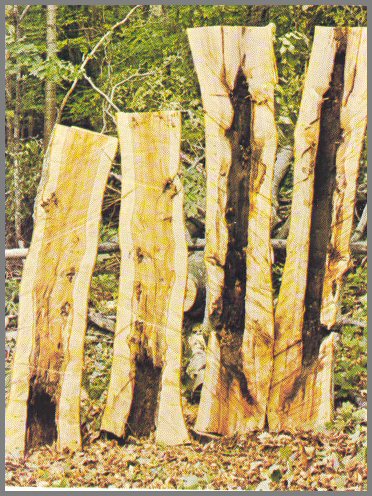
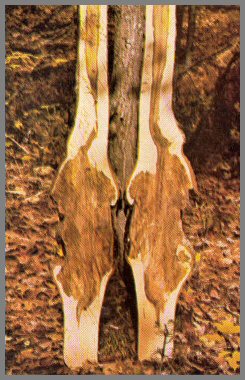
FIGURE 41.-EUTYPELLA CANKERS ON SUGAR MAPLE.
Eutypella parasitica caused the cankers on these two sugar maples. Decay by other fungi followed. The fruit body of E. parasitica is minute and black, and has a neck about 1/4 inch long.
Dissection shows that the cankers and the unhealed stubs have made these two trees worthless. However, the defect associated with the cankers alone is very slight. Stems often break at canker sites.
Page 59
FIGURE 42.-0THER CANKERS ON SUGAR MAPLE.

A canker of unknown origin on a sugar maple. Such cankers are very hard. They are usually found at branch stubs. They are similar to those caused by Polyporus glomeratus, except that no sterile conk is present.

Dissection shows that very little defect is associated with this canker. When in doubt about a canker like this, cut it open with an ax: if you find no sterile conk, the defect will be limited to the canker area.
Page 60
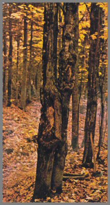
FIGURE 43.-STEM SWELLINGS.
This sugar maple tree had two healed swollen areas near the base. Dissection shows very little defect. When such areas are healed tightly, very little defect is associated with them. Note that the central column of discoloration narrows abruptly toward the base. This kind of stem swelling is common in some areas.
Page 61
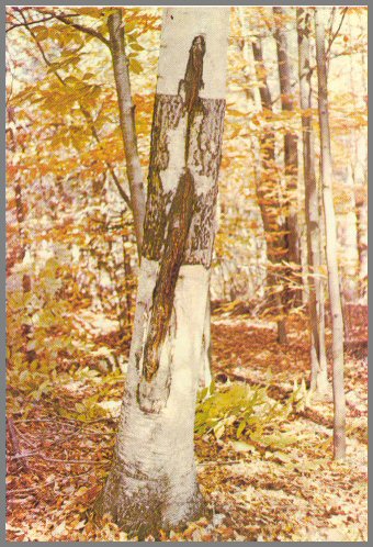
FIGURE 44.-SUNKEN STEM CANKER.
This sunken stem canker on a paper birch is caused by Fomes igniarius
var. laevigatus. These sunken cankers are most common on mature and
overmature paper birch and yellow birch trees. They indicate advanced
decay-especially on trees with large poorly healed stubs. The fruit body
resembles a flattened fruit body of F. igniarius.
Page 62
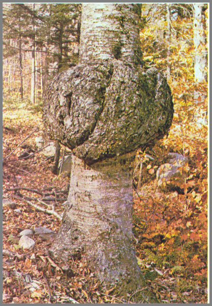
FIGURE 45.-STEM BURL
A stem burl on an ash tree. Burls and large swollen areas - often
called tumors - can be found on all northern hardwood species. The bark is
usually intact.
Dissection shows very little defect is associated with burls like this-unless
the bark is broken; then decay is often present. Wavy grain is usually
found a short distance above and below these abnormal growths. The cause
of these burls is not known.
Page 63
FIGURE 46.-FUSIFORM SWELLING.
Fusiform (spindle-
shaped) swelling on the stem of a yellow birch. The bark is rough, thick, corky, and dark on these swollen areas. Swellings like this are also found on other species of northern hardwoods.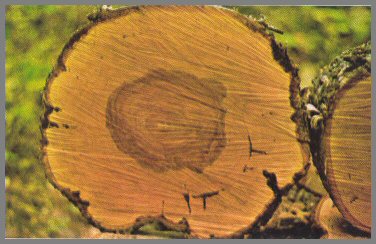
Dissection shows very little defect, except hard dark spots in the wood and some wavy grain a short distance above and below the swelling. However, if an ooze is found on the swollen area, you can expect extensive discoloration. The cause of such swelling is not known but a disease of the outer bark after it has been wounded is suspected.
Page 64
FIGURE 47.-CORKY BARK.
This sugar maple has a swollen area, with rough corky bark. The wood is not exposed as in Nectria cankers. Bark insects are common in the corky bark. Such trees are often found in clusters.
Dissection shows that only slight discoloration is associated with the swollen area under the corky bark. The wood is dense and hard, and often separations occur between some of the growth rings.
Page 65
FIGURE 48.-LARGE BURLS.
Large burls can be found on all northern hardwood species. This one is on a yellow birch. Very little defect is associated with these large burls as long as the bark is tight. Trees with burls like this are often found in clusters.
Page 66
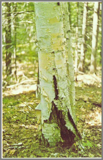
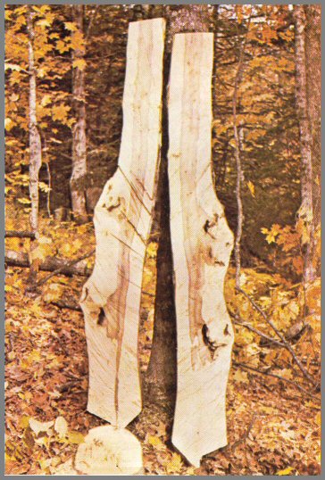
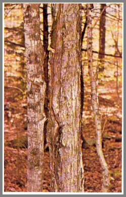
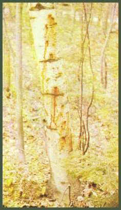
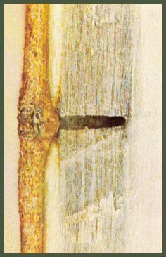
FIGURE 49. - SEAM OR FROST CRACK ON PAPER BIRCH.
The black band on this paper birch marks where cracks formed above and below a wound. The white bark usually falls away from such areas. (This should not be confused with the black bands that form after bark has been stripped off.) The cracks have lips that open in winter and close in summer. Seams or frost cracks often begin at wounds and branch stubs. Discoloration is associated with all cracks, and sometimes decay is too.
Page 67
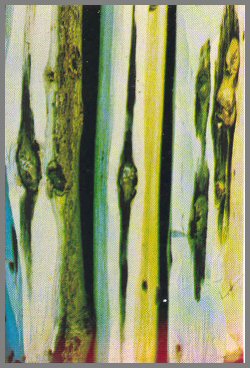
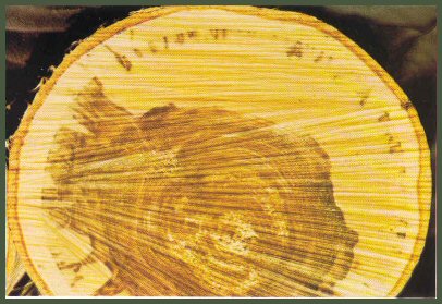
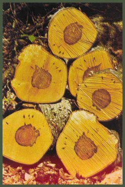
FIGURE 50.-SEAM OR FROST CRACK ON RED MAPLE.
This red maple has a large
seam or frost crack, bearing a fungus fruit body. Discoloration and decay is usually associated with stem cracks like this. The open lips on this crack indicate extensive defect.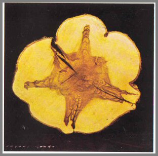
Cross-section of a red maple stem with five frost cracks shows the pattern of defect. Though the defect often is only a slight discoloration, the pattern makes it difficult to saw out a board free of discoloration.
Page 68
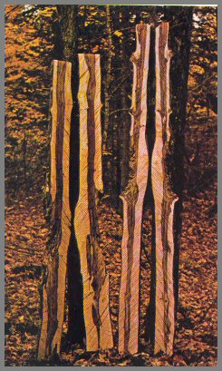
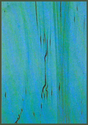
FIGURE 51.-SAPSUCKER INJURY.
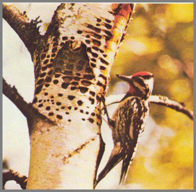
The yellow-bellied sapsucker, which drills holes in living trees, causes considerable damage in the forest.
This section of a yellow birch stem shows typical discolorations from l-year- old sapsucker wounds. The streaks may be a few inches to more than a foot long. In cross-section they create islands of discoloration.
Page 69
Figure 51, Continued
This shelled-out section of paper birch stem illustrates the ring shake that may form in living trees where sapsuckers have caused severe damage. Some wood shakes apart later after the boards have been sawed and dried. Drill holes made by the sapsucker can be recognized in the bark after more than 50 years.
This black band on a paper birch bolt is typical of severe injury caused by sapsuckers. Usually seen high on the tree, these bands can be found on all northern hardwood species.
Page 70
FIGURE 52.-SQUIRREL INJURY.
This wound on a small sugar maple was caused by a red squirrel. Red squirrels bite young trees in the spring to start sap flows, which they drink. The teeth marks can still be seen on the wound surface.
Dissection of a small red maple shows the discoloration caused by injuries inflicted by a red squirrel.
Page 71
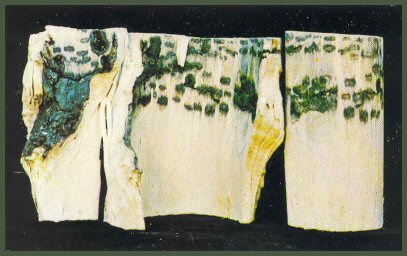
FIGURE 53.-AMBROSIA BEETLES.
The bleeding from this paper birch is from holes made in the bark lenticels by ambrosia beetles. Small bleeding spots like these can be found on all northern hardwood species; and dead spots in the bark are usually associated with them. Bleeding spots may indicate early canker formation.
A hole made in a paper birch by an ambrosia beetle. The discoloration extends several inches above and below the hole. Small holes in bark lenticels signal this kind of defect. Such trees should not be used for veneer.
Page 72
This cross-section of a paper birch bolt shows the islands of discoloration due to holes made by ambrosia beetles. The islands in some areas merge with the central column of discoloration.
These bolt ends show effects of a light infestation by ambrosia beetles.
Page 73
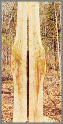
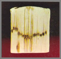
FIGURE 54.-CAMBIUM MINER (PHYTOBIA SP.).
This piece of paper birch veneer shows damage due to the cambium miner. Defects like these are sometimes called worm tracks. The injury to the tree is slight, but the damage to the wood is great. The injury can be seen as soon as the tree is cut. In cross - section the insect galleries appear as brown to orange-brown pencil marks about 1/16 to 1/8 inch long within the growth rings. If the marks are concentrated in the center of the tree, little damage is done. But if they are scattered throughout the stem, it is ruined for veneer. Cambium miner marks occur in all the northern hardwood species. In the maples they are brown.
Page 74
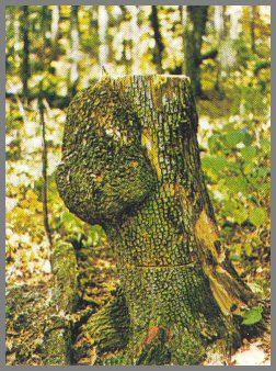
FIGURE 55.-PORCUPINE INJURY.
The wound at the base of this beech tree was caused by porcupines. They attack all the northern hardwood species. The teeth marks can be seen even after the wounds are very old. These wounds should be regarded like any other kind of mechanical wounds. Discoloration is commonly associated with them; and decay is associated with old wounds that have dark splintery surfaces. Beavers cause similar injury.
Page 75