Page 1-25
[Note from the web master. The word heal may be used here to describe the closure of a wound. Dr. Shigo now calls it closure and not healing. That and other terms can be found at www.treedictionary.com for your use. John A. Keslick , Jr.]
HOW CAN YOU TELL?
H
ow CAN YOU TELL, by looking at a living tree, what quality it has? One tree may be rotten at the core yet still may contain a large volume of high-quality wood. Another may be thoroughly sound from pith to bark yet may be so damaged by minute discolorations that it is worthless for high-quality products. How can you tell?Page 1
By dissecting living trees and studying the organisms that
infect them, we now know that discoloration and decay develop in certain
definite patterns. And the patterns of discoloration and decay within the
tree can be predicted from external signs.
Discoloration and decay are the most serious defects of
northern hardwood trees. In speaking of defect, we must distinguish
between injury and damage. Injury harms the tree: damage lowers the
quality of the wood. For example, a disease like vascular wilt may kill
the tree but do no damage to the wood. But an insect like the cambium
miner may do very little harm to the tree yet do great damage to the wood.
The unseen damage done to a tree is important in the
economics of forestry. Every operation in growing a tree, harvesting it,
and converting it into products costs time and money. And after all the
time and money have been spent on a tree, the product made from it may prove to
be not worth the effort; and the tree might have been used more profitably for
some other product that does not require high-quality wood.
The increased use of veneer offers an illustration. A
veneer log brings top prices. But its actual value may not become apparent
till it is put on a lathe and peeled. A log that looks very good, and
sound to the core, may have minute streaks of discoloration scattered all
through it, so that all the veneer it produces is badly streaked with defects.
On the other hand, a log that has a rotten core surrounded by clear wood may
produce the highest quality of veneer.
So it is not so important how much discoloration and decay a
tree has, but where these defects are in a tree. The pattern of the
discoloration and decay-that's the important thing.
STUDY METHODS & MATERIALS
This guide is based primarily on the findings from a series of studies made by the senior author in northern New England. Begun in 1959, these studies are being continued in efforts to clarify further our understanding of the discoloration and decay processes.
Page 2
The Species
The northern hardwoods make up one of the major forest
types of northern New England. They are American beech (Fagus
grandifalia Ehrh.), paper birch (Betula papyrifera Marsh.), yellow
birch (B. alleghaniensis Britt.), sugar maple (Acer saccharum
Marsh.), red maple (A. rubrum L.), and white ash (Fraxinus americana
L.).
Most of the sample trees grew in the White Mountain National
Forest in New Hampshire.
The resource of northern hardwoods is plentiful today, but it
contains an overabundance of poor-quality trees. Foresters repeatedly ask
two questions about the northern hardwoods: What can we do to assure future
crops of high-quality trees? And how can we make the most of what we have
now?
The Methods
A new large-scale method of dissecting living
trees was used in these studies, to get at the defects inside the tree.
This might have seemed an obvious thing to try. Yet large-scale dissection
had not been tried on trees-at least not in quite this way.
True, pathologists had studied decay in wood and had isolated
and identified fungi. Studies had been made of the cut ends of logs.
And wood defects had been studied on freshly cut boards at the sawmill.
But logs taper, and many of them are not straight; and though the surfaces of
lumber cut at a sawmill reveal discoloration and decay, they do not reveal the
complete patterns.
The development of the gasoline-powered chain saw since World
War II made possible this method of dissection. By using a portable chain
saw, it was possible to go into the forest and work on living trees. One
could fell a tree and cut it into bolts and disks at once and rather quickly-a
tedious and discouraging job with hand saws. Moreover, it was now possible
to begin at one end of a tree stem and slice it down through the middle,
following the natural curve of the stem. And it was possible to cut at any
spot and any angle, and to lay open any particular area of a tree for study.
After a tree had been cut open, the defects were
systematically
Page 3
mapped. Then, from specimens of wood taken into the laboratory, chips 1
x 0.3 cm. were cut with a gouge from clear wood, discolored wood, and decayed
wood, and from the zones between -six chips in each series, at intervals up and
down the tree stem. These chips were cultured on agar plates. Every
organism found was recorded, and most were identified.
Scope of the Studies
During these studies more than 3,000 trees were dissected,
and some 100,000 isolations were made to identify organisms. More than
10,000 photographs-both black-and-white and color -were taken to record the
patterns of discoloration and decay in the freshly dissected trees. From
these, 100 color photographs have been selected to illustrate this guide.
GENERAL RESULTS
As dissection of the living trees disclosed general patterns of discoloration and decay, a clear understanding about three aspects of the discoloration and decay processes emerged.
A Succession of Organisms
Decay in a tree had been thought to center about three simple
events-a tree is injured: a fungus enters: decay begins. But our studies
showed that a complex succession of events must take place before decay can
begin. It works roughly like this-a tree is injured: the tree reacts:
chemical changes take place in the wood: the wood discolors: bacteria and
non-decay fungi become active: the wood discolors further: decay fungi infect:
decay begins.
The process is irreversible. And it cannot be
shortcut. Decay does not begin until all the other events in the
succession have taken place. Decay does not begin until the wood has been
discolored. And the succession may stop at any stage: the wood may become
discolored but may not decay.
A Consistent Pattern
The discoloration and decay in a northern hardwood tree
take a definite pattern. They form a column in the tree, related
Page 4
to the location of the injury. This column can spread up and down inside the tree, but it seldom spreads outward. The diameter of the column of defect is no larger than the tree was at the time of injury. In effect, growth of the tree after the injury forms a pipe of healthy new wood, and the discoloration and decay are held within this pipe.
Discoloration not Heartwood
In some tree species, like black cherry and walnut, true
heartwood forms as a result of chemical change that follows normal aging
processes in the wood cells. But in northern hardwoods the darker wood is
not true heartwood in this sense: it is a result of discoloration processes
initiated by injury.
THE PROCESSES OF DISCOLORATION AND DECAY
The development of discoloration and decay in the wood of a living northern hardwood tree is a complex continuum of events that merge and overlap in time and place. Several columns of discoloration and decay may be present in various stages of development at the same time and place. It is difficult to say where one stage ends and another begins. But for the sake of convenience in describing the processes, we divide them into three broad stages.
Stage 1
The process begins with an injury to the tree. A
branch may die or break off; insects or birds or animals may attack the tree; a
fire may burn the base; or a logging machine may scrape it in passing.
Some cells are killed by the injury, and others may be injured to some degree.
The injured cells are exposed to the air. At this time gases and moisture
can pass out of the tree, and air and moisture can pass into the tree.
These changes start chemical processes in the wood cells about the wound.
Discoloration results. It may be due to the materials
formed by the chemical processes, or to the darkening of cellular material
Page 5
as a result of exposure to the air. Sometimes the discoloration may be
a bleaching rather than a darkening. Microorganisms are not involved.
These early discolorations do not alter the strength of the
wood. And the process may stop right here. It depends on the
severity of the wound and the vigor of the tree. But the discoloration may
advance inward toward the pith, and around the tree.
Then in time a new growth ring forms. The first
cells in this ring are different from the cells that are usually produced.
They act as a barrier to the discoloration process. The discoloration
seldom moves into the new cells. Instead, it moves up and down the stem
within the pipe of barrier cells-but not outward into the new tissues.
The extent of discoloration depends on the vigor of the tree,
the severity of the wound, and on time. Discoloration advances only as
long as the wound is open. Thus the entire cylinder of wood present when
the tree was wounded may not become discolored. Meanwhile the tree
continues to form new growth rings that are free of discoloration.
Stage 2
Stage 2 begins when microorganisms infect. As soon as the tree is
wounded, many different organisms begin to grow on the wound surface. They
compete; and many do not survive. Of those that survive, only a few types
are able to begin growing into the wood through the wound-only those that can
thrive in the discolored wood.
These first microorganisms to invade the tree-the pioneers-
are bacteria and fungi. The fungi are non-Hymenomycetes: they do not cause
decay. These pioneers infect only the cells that have been altered by the
chemical processes, so the new tissues formed after the injury remain free of
infection.
The infected cells are further altered by the pioneer
microorganisms. The discoloration may increase; the cells become more
moist; pH (acidity) and mineral content of the cells rises; and certain parts of
the cell walls may be eroded. The wood
Page 6
affected at this stage is called wetwood, redheart, or blackheart, The process may stop at this stage.
Stage 3
Stage 3 begins when decay fungi (Hymenomycetes) become active and begin to
digest the cell walls. These fungi affect only those tissues that first
have been altered by chemical processes and then by the pioneer organisms.
The new growth of wood that continues to form remains free of infection.
The decay process continues as long as the wound remains
open. Many species of microorganisms may interact until the wood is
completely decomposed. The succession of organisms does not stop when the
first decay fungus enters. It stops when the tissues are completely
digested.
The advancing decay column is often separated from the new
white wood by a band of discolored wood. In some species the margin of
this band may be bleached. The pioneer organisms remain in this
band. Later, as decay continues, the decay fungi slowly digest this
discolored band; and only a hard black rim then separates the by-then hollow
core from the healthy white wood. This rim forms first near the wound.
To this point the process may take 40 to 50 years.
Healthy white blemish-free new wood will surround the hollow core, unless other
wounds have been inflicted meanwhile.
We want to emphasize this point: the processes need not go
through to completion. Healing of a wound, antagonisms among organisms,
unfavorable environment, and other forces may cause the processes to abate in
any stage.
Importance of Time
What we have described above is a continuum of events
that follows a single wound at one period in the life of the tree. But a
forest tree is apt to be injured a number of times. The same processes
take place each time. And the discoloration and decay processes that
follow a new wound are not affected by the effects of older wounds.
Page 7
THE PATTERNS OF DISCOLORATION AND DECAY
A cross-section of an ideally healthy northern hardwood tree shows a pencil-thin cylinder of pith in the center of the stem, surrounded by unblemished white wood. But what you see in most trees is a core of darker wood, of varying diameter, surrounded by white wood. The pattern of the discolored wood will depend upon what has happened to the tree: when the branches died, how fast it healed its wounds; what logging wounds it had; and how much injury was done to it by insects, fire, birds, animals, or other agents.
Central Column
The most typical pattern of discoloration is a column of
discolored wood extending up and down in the center of the tree. This
discolored wood is always associated with some injury to the tree, like a broken
branch or stem. Where decay occurs, it occurs within this column of
discoloration.
In northern hardwoods, this central column of darker wood is
not true heartwood-though it is often called heartwood. It does not
increase in diameter as the tree grows. The discoloration is due to
changes in the wood brought about by processes that are begun by wounds or other
injuries..
True heartwood does occur in some hardwood species such as
black walnut, cherry, and the oaks. This darker wood is due to processes
associated with normal aging of the tree. As the tree grows, the column of
darker wood increases in diameter. The cylinder of true heartwood extends
rather uniformly throughout the entire tree, from the base up into the branches.
Columns of discolored and decayed wood, caused by injury to
the tree, can form within the column of true heartwood. These columns of
discolored and decayed wood do not increase in size as the heartwood does.
Of course in a tree like black walnut the difference between true heartwood and
discolored wood is hard to distinguish.
The differences between true heartwood and discolored wood
can be tabulated roughly this way:
Page 8
Click here for comparison chart.
Multiple Columns
When a tree is injured at different times, multiple
columns of discolored wood develop. The multiple columns can be seen most easily
on the ends of logs, where they take on a concentric pattern or a cloud-like
pattern. Very careful dissections are necessary to trace each column to
its source.
If two injuries to branch stubs occur at about the same time,
the two columns of defect may join to appear like one column.
Page 9
But if wounds occur at different times, the processes that
result in discoloration and decay begin and develop independently for each
wound.
If a cylinder of discoloration is already present in the
stem, a later wound does not affect it. The discoloration from the new
wound forms around or beside the old column of discoloration. It may
completely envelop the old column, like a pipe sliding down over a smaller pipe.
Or it may develop alongside the old column in the shape of a crescent or half
moon. It may be wedge- shaped in cross-section.
Major injuries to the tree-such as broken stems and branches
-tend to discolor the entire core of the tree that was present at the time the
injury occurred. Minor injuries such as small insect wounds may cause only
localized streaks of discoloration-islands of defect within sound white wood.
Because each column of defect is separate in both time and
cause, the discoloration and decay processes may be more advanced in one column
than in another. Decay may form in one crescent-shaped column while other
adjacent columns may be discolored but still sound. Sometimes columns of
defect enclose areas of clear wood. For example, a tree may have a sound
center surrounded by a ring of decayed wood.
When the wood begins to dry after the tree has been cut,
checks often form between the boundaries of the defect columns. The checks
often follow the growth rings-ring shake. When the defect column is a
complete cylinder in cross-section, the shake may develop in a complete circle
along the growth ring. Because the defect columns may be in different
stages of the discoloration and decay process, the stresses that develop in
drying are not uniform; so checking may occur in many different patterns.
For example, this is common in sugar maple when branch stub
wounds and borer wounds occur in the same place on the stem. Honeycombing
or tissue collapse may result in the moist discolored tissues. And because
the beetles wound the wood in a diagonal pattern, crossing many growth rings, a
jagged column of defect sometimes forms that checks in all directions when the
wood dries.
Page 10
Defects under Cankers
The defects that form under cankers are somewhat
different. A canker tends to produce a localized defect rather than a
column of discoloration and decay.
When a canker develops about a wound, the tree usually
already has a central column of discoloration and decay. The defect from a
canker usually forms beneath the canker, in the wood between the central column
of discoloration and the bark surface area where the canker forms. The
defect from a canker usually does not spread around the entire stem: it lies
immediately beneath the canker.
Mineral Streak and Stain Wounds also begin the processes that
result in the discolorations that are commonly called mineral streaks. The
accumulations of salts in such discolorations may be due to the evaporation of
liquids that are drawn to the exposed wound, as in a wick action. Because
fungi are the agents most consistently associated with wounds does not mean that
other organisms, especially bacteria, do not also alter the wood. And
viruses can also cause discolorations; so they cannot be ruled out as agents
inciting stain.
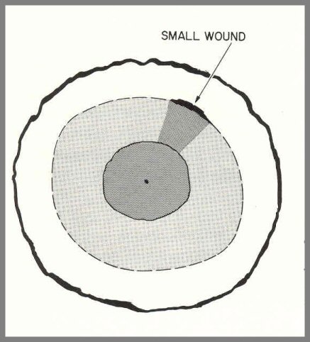
Above: Even though an entire central column may not be discolored by a small wound, the whole central column may take on a slightly darker hue.
Page 11
Other Patterns
The wounds caused by sapsuckers, fire, and other agents have some typical
patterns, but the course of discoloration and decay follows the same patterns
described above.
Sometimes, when a wound is small and not severe, the discoloration
affects a small area rather than a complete new column; yet the new central
column takes on a slightly darker hue -usually light pink.
EFFECTS ON DIFFERENT SPECIES
All the northern hardwood species do not react exactly the same way to injuries. Some notes about each of these species follow.
Sugar Maple
Of all the northern hardwoods, the most resistant to discoloration
and decay is sugar maple. Yet many sugar maple trees are very defective.
This is because some agents of injury seem to prefer sugar maple to the other
species: for example, sap- suckers, cambium miners, squirrels, and-most
important-the sugar maple borer. Discolorations associated with wounds of
sugar maple are often called mineral streaks. Of the fungi that cause
decay in sugar maple, the most important are Fomes connatus, F. igniarius,
and Polyporus glomeratus.
Red Maple
In general, red maple is considered to be very susceptible to
defect. Discoloration and decay advance much faster in red maple than they
do in sugar maple. The central columns of discoloration are usually due to
branch stubs, most of them between 4 and 10 feet up on the stem.
Sprout clumps are common in red maple, and they present some
serious problems. Branch stubs on sprout stems of red maple cause more
defect than the old parent stumps cause.
Many fungi cause decay in red maple, but the principal one is
Polyporus glomeratus.
Page 12
Yellow Birch
Discolorations and decays advance in this species fastest of all. Large low branch stubs - usually not well healed-are most common in yellow birch. In older trees, top breakage accounts for wide columns of discoloration. Yellow birch is one of the favorite feeding trees for sapsuckers. The most common fungi that cause decay in yellow birch are Poria obliqua, Pholiota species, and Fomes igniarius. F. igniarius var. laevigatus causes cankers on overmature trees.
Paper Birch
Paper birch reacts like yellow birch in most ways, except
that discoloration and decay do not advance so rapidly, and large low branch
stubs are not so common. Advanced decay is like that in yellow birch.
The cambium miner is more common on paper birch, and so is the ambrosia beetle.
Paper birch is not a favorite feeding tree for sapsuckers; but they do attack
it, and heavy attacks cause black bands on the stem. The same decay fungi
that attack yellow birch also attack paper birch.
Beech
The beech bark disease is the most important disease of
beech. It has decimated beech in many areas.
One special feature of beech is that its base is vigorous,
and wounds near the base rarely cause much damage to the roots. The
notable exceptions are the defects caused by Fomes applanatus and
Armillaria mellea. Because of its vigorous base, a beech tree with a
defective bole may stay alive for a long time.
Ants sometimes infest wounds on beech trees, and their
activities increase the defect by keeping the wound open. These insects
are rarely found on other northern hardwoods. Branches from dormant buds
on the stem may cause small defects when they die.
Ash
In ash trees, most of the defect comes from the top downward.
Poorly healed stubs in the crown, and broken tops, should be considered
important in this species. The principal decay
Page 13
fungus of ash is Fomes fraxinophilus, which affects the upper stem. Frost cracks are common on ash in some areas.
STUBS WITHIN THE STEM
How to Estimate Size, Angle, and Depth
The size and position of a branch stub buried in the wood
inside a tree can be estimated roughly from the scars on the bark. And
from this in turn you can estimate the size of the defect column and the amount
of clear wood outside it. Much of the information on this subject comes
from research done in Europe.
Two types of bark scars provide the clues. One, called
a chinese beard, is like two drooping moustaches. The other, like a mouth
under the moustaches, is the more or less round scar that marks where the stub
was.
The chinese beard forms this way: as a branch grows on the
tree stem, a layer of thick-walled cells forms in the axil above the branch.
The bark becomes roughened over this ridge of thick-walled cells. In some
species, like paper birch, the bark also turns dark here.
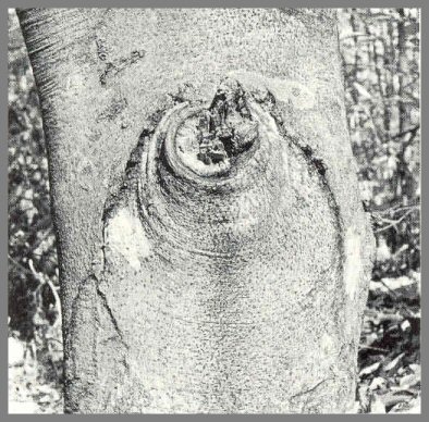
Above: Stub scar and chinese beard on a beech tree.
Page 14
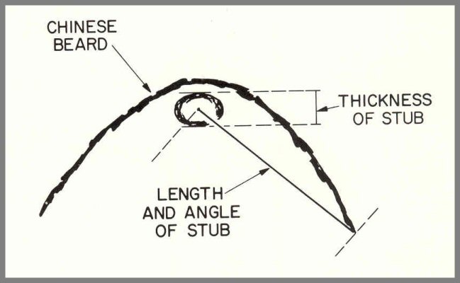
Above: How length and angle of stub is estimated from the stub scar and chinese beard.
This scar can be used to estimate both the length of the stub
inside the tree, and the angle at which the stub
joins the vertical axis of the tree center. From the center of the stub
scar, draw a line to the bottom end of the chinese beard. This equals the
length of the stub and shows the angle at which it lies.
The thickness of the stub can be estimated directly from the
stub scar. Assume that the branch that broke off to form the stub was
practically round. The height of the stub scar shows the size.
If the stub scar is about 2 inches high, the stub is about 2 inches in diameter.
How far inside the tree the stub is buried can be estimated
from the shape of the stub scar. As a tree adds new growth rings,
the growth at anyone spot on the stem is outward, not upward. The height
of the stub scar remains the same. But as the new growth is added, the
bark is pushed outward, and the scar spreads at the sides, gradually changing in
shape from a circle to an ellipse.
The scar's ratio of height to width indicates how far the
stub extends from the pith center of the tree toward the bark; and this tells
conversely how much new wood has formed over the stub.
Page 15
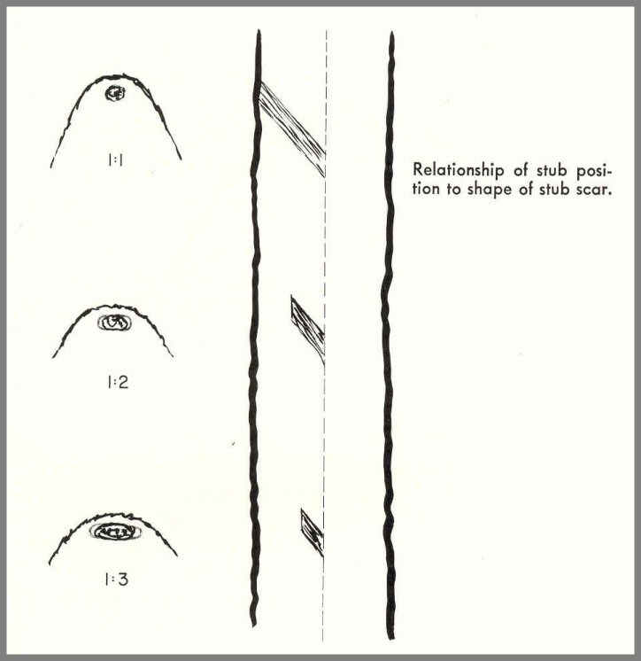
For example, if the stub scar is round, it has a ratio of
1 : 1. This means that the end of the stub lies just at the bark surface
-an unhealed stub-and no new growth covers it. If the scar is twice as
wide as it is high, the ratio is 1 : 2. This means that the stub extends 1/2 the
distance between the pith center and the bark, and that the other 1/2 is white
wood. And if the ratio is 1 : 3, the stub extends 1/3 the distance between
pith center and bark. And so on.
This rough formula can be applied to all the northern
hardwood trees. As time passes, use of this formula becomes more
difficult, especially on the maples.
Page 16
PHOTO GUIDE
These photographs illustrate all the major defects of northern hardwoods, as we now recognize them, and the fruit bodies of the most important decay fungi.
Page 17
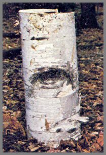
FIGURE 1. - LOW BRANCH STUB.
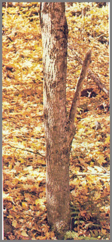
This sugar maple (above) has a low stub, but the base is free of
wounds. The long stub of hard wood shows that the stub has not been dead
long, and that the discoloration processes have not had time to progress to an
advanced stage.
Page 18
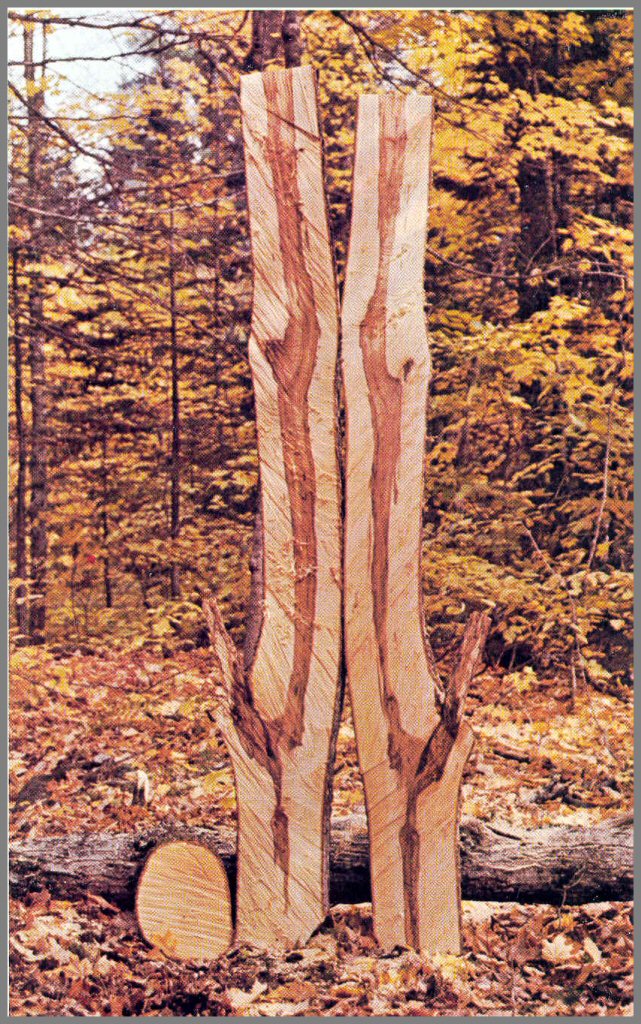
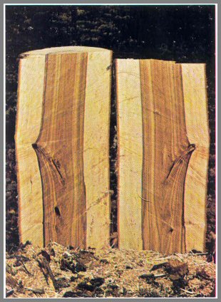
Dissection (above) reveals the pattern of the discoloration. The wood at the base is clear. The column of discoloration from the large stub dwindles toward the base, but joins above with a wider column of discoloration from an older stub above, where the processes are more advanced. The wood formed after the stubs died remains free of discoloration. These discolored tissues are not heartwood.
Page 19
FIGURE 2. - LOW BRANCH STUB DECAYED
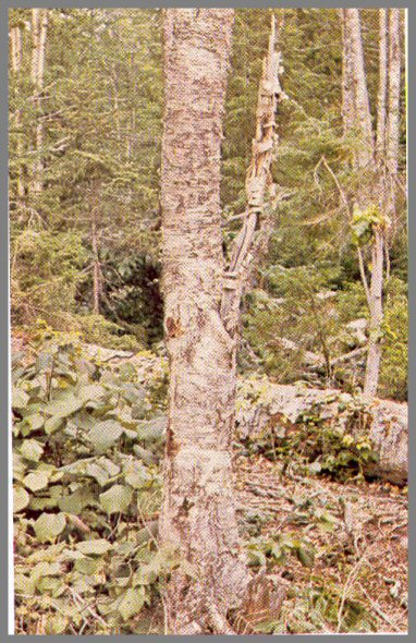
This yellow birch (above) has a low stub, and the callus ridge and advanced decay show that the stub has been dead a long time. Roughened bark at the base indicates basal wounds and root wounds typical of injury caused by logging equipment.
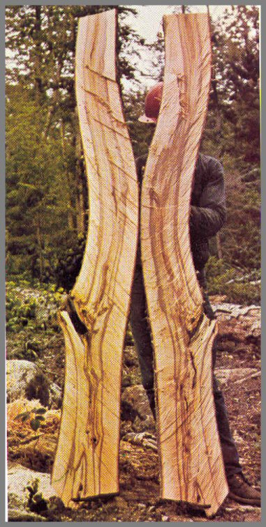
Dissection (above) reveals discoloration and decay originating from both the basal wounds and the stubs above. The base is both discolored and decayed. The dark lines within the decay at the base indicate the limits of earlier columns of discoloration. The diameter of the widest column indicates the diameter of the tree when - the large branch died.
Page 20
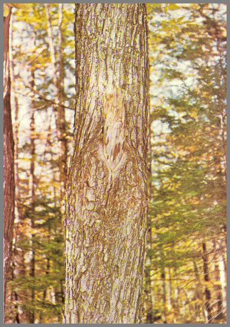
FIGURE 3. - HIGH BRANCH STUB, UNHEALED
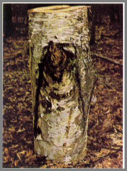
This paper birch bolt (above) contains a large unhealed stub from high on the tree. The decayed stub indicates advanced discoloration and some decay. The dark drooping lines of the bark scar - chinese beard - indicate the angle and length of the stub inside the stem.
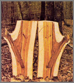
The discolored wood associated with the stub is redheart - very wet and dark, and - beginning to decay. The column of defect from the stub does not enter the older central columns of defect, but extends up and down alongside them. Multiple columns of defect like this are common.
Page 21
FIGURE 4. - HIGH BRANCH STUB, HEALED
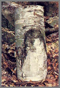
This healed stub wound on a paper birch illustrates how the
position of the stub inside the tree can be estimated. The dark line of
the Chinese beard shows the length and angle of the stub. The height of
the stub scar tells the diameter of the stub.
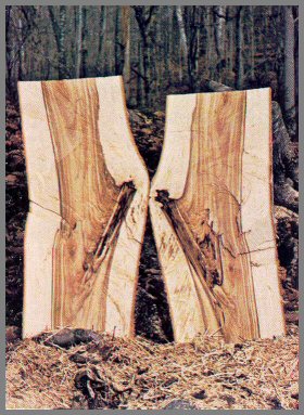
Dissection shows the relationship. The length, angle, size,
and depth of the stub are approximately as indicated by the external signs.
Note that the stub healed before the last bit broke off, leaving a pinched-off
piece; this affects the accuracy of the formula. In this bolt the
discolored wood is as sound as the white wood; it is not moist (thus not
redheart) , and no organisms have invaded to alter the wood.
Page 22
FIGURE 5. - HIGH SMALL BRANCH STUB, HEALED
Full view of a small well- healed stub from high on a paper birch tree.
The ellipse form of the stub scar (a 1 : 2 ratio) shows that the stub is buried
about halfway between the tree center and the bark. Small, high, well-healed
stubs like this indicate discolor- ation, but not decay.
Dissection reveals that the discolored wood in the stem is sound. The
small stub was associated with a column of discoloration that formed after the
branch died, when the tree was about 8 inches in diameter at this point. A
central column of discoloration had formed earlier after branches died when the
tree was about 4 inches in diameter. This is shown by the light-colored
boundary streak in the discolored core. As other larger stubs died above,
other columns of discoloration formed, enveloping the older and smaller columns.
Page 23
FIGURE 6. - BRANCH STUBS IN SMALL STEMS
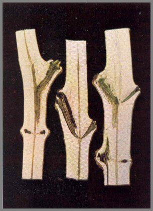
Dissection of these small red maple stems shows that
discolorations begin early in the life of the tree. At this age, poorly
healed stubs give invading organisms the advantage. A young tree that
tends to heal branch wounds slowly is a poor risk for a crop tree.
Page 24
FIGURE 7, - HYPOXYLON RUBIGINOSUM ON STUB
The red-brown patches on this large low red maple stub are fungus material that
contains fruit bodies of Hypoxylon rubiginosum, a pioneer invader of
wood. Its presence indicates that discoloration is advanced and decay is
beginning. Trees that have several large low stubs like this usually have
large central cores of defect. Large cracks may form under such stubs; and
then the discoloration and decay processes go faster.
H. rubiginosum is one of the few non-Hymenomycetes that cause decay as well as discoloration. I t is one of the most aggressive fungi in the forest. It infects both living trees and slash.
Page 25